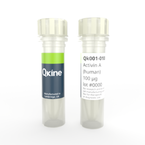
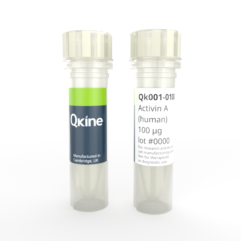
 Recombinant human activin A protein (Qk001)
Recombinant human activin A protein (Qk001)
Price range: £250.00 through £3,190.00
Activin A is a TGF-β family growth factor regulating embryonic development, cell proliferation, differentiation, and immune responses. Activin A is frequently used to maintain pluripotency in induced pluripotent and embryonic stem cell cultures. It is also used in many stem cell differentiation protocols, including endoderm lineage differentiation and further maturation into hepatocyte and pancreatic cells.
Recombinant human activin A protein is a high-purity mature bioactive dimer of 26 kDa. It is animal origin-free (AOF), carrier protein-free, and tag-free to ensure its purity with exceptional lot-to-lot consistency. This protein has been rigorously benchmarked against other commercial sources and extensively validated for highly reproducible stem cell culture. Qkine founder Marko Hyvönen developed the protocol for making high-purity activin A, and this protein is used at the Cambridge Stem Cell Institute.
High purity mature bioactive dimer rigorously benchmarked against other commercial sources.
Qkine activin A is also available as cell therapy grade with extended quality testing and documentation – Qk001-CTG
In stock
Orders are typically shipped same or next day (except Friday).
Easy world-wide ordering, direct or through our distributors.
Price range: £250.00 through £3,190.00
Buy online with secure credit card or purchase order. For any questions, please email orders@qkine.com
Summary:
- Bioactive mature domain of recombinant human activin A protein, residues 311-426 (Uniprot: P08476)
- 26 kDa (dimer)
>98%, by SDS-PAGE quantitative densitometry
Expressed in E. coli
Animal origin-free (AOF) and carrier protein-free
Manufactured in our Cambridge, UK laboratories
Lyophilized from acetonitrile, TFA
- Resuspend in 10 mM HCl (Reconstitution solution A) at >50 µg/ml, add carrier protein if desired, prepare single-use aliquots and store frozen at -20 °C (short-term) or -80 °C (long-term)
Featured applications:
Induced pluripotent and embryonic stem cell differentiation and maintenance
iPSC-derived mesoderm differentiation
Differentiation of iPSC into endoderm
Chemically defined media for organoid culture
- Recombinant human FGF-2 (145 aa) protein (Qk025)
- Recombinant human BMP-4 protein (Qk038)
- Recombinant human EGF protein (Qk011)
- Recombinant human BMP-2 protein (Qk007)
- Recombinant human TGF-β1 PLUS™ protein (Qk010)
- Recombinant human FGF-2 (154 aa) protein (Qk027)
- Recombinant human IGF-1 protein (Qk047)
- Recombinant human IGF-1 LR3 protein (Qk041)
- Recombinant human NRG-1 protein (Qk045)
- Recombinant human OSM protein (Qk049)
- Recombinant porcine EGF protein (Qk064)
- Recombinant mouse EGF protein (Qk066)
- Recombinant bovine PDGF-BB protein (Qk079)
- Recombinant chicken/duck FGF-2 (145 aa) protein (Qk030)
- Recombinant zebrafish FGF-2 protein (Qk002)
- QkPlate© human vitronectin coated 6 well plate (Qk2001)
- Recombinant human nodal protein (Qk029)
- Recombinant human activin A PLUS™ protein (Qk005)
- Recombinant human follistatin-resistant activin A (FRACTA) protein (Qk035)
- Tri-lineage differentiation kit (Qk516)
- StemBeads® Qkine activin A
- Recombinant human activin A protein (Qk001-CTG)
- Recombinant mouse/rat activin A protein (Qk012)
- Recombinant mouse/rat activin A PLUS™ protein (Qk014)
- Serum-free media optimization growth factor discovery kit (Qk505)
- Endoderm differentiation kit (Qk513)
- Mesoderm differentiation kit (Qk514)
- Recombinant human activin B protein (Qk024)

Activin A activity was determined using the activin-responsive firefly luciferase reporter assay in transiently transfected HEK293T cells. Cells were treated in triplicate with a serial dilution of activin A. Firefly luciferase activity was measured and normalized. EC50 = 11.9 pM (309.4 pg/ml). Data from Qk001 lot #104276.
Activin A migrates as a single band at 24 kDa in non-reducing (NR) and 13 kDa as a single monomeric species upon reduction (R). No contaminating protein bands are visible. Purified recombinant protein (7 µg) was resolved using 15% w/v SDS-PAGE in reduced (+β-mercaptothanol, R) and non-reduced conditions (NR) and stained with Coomassie Brilliant Blue R250. Data from Qk001 lot #011.

Further quality assays
Mass spectrometry: single species with expected mass
Recovery from stock vial: >95%
Endotoxin: <0.05 EU/μg protein
We are a company founded and run by scientists to provide a service and support innovation in stem cell biology and regenerative medicine. All our products are exceptionally high purity, with complete characterisation and bioactivity analysis on every lot.

Qkine activin A was as bioactive as activin A protein from an alternative supplier. Activin A activity was determined using the activin-responsive firefly luciferase reporter assay in transiently transfected HEK293T cells. Cells were treated in triplicate with a serial dilution of Qkine activin A (Qk001, green) or alternative activin A (Supplier B, black). Firefly luciferase activity was measured and normalized. Data from Qk001 lot #204536 EC50 = 16 pM, Supplier B EC50 = 12 pM.

Qkine activin A had consistent bioactivity over 3 independently manufactured lots. Activin A activity was determined using the activin-responsive firefly luciferase reporter assay in transiently transfected HEK293T cells. Cells were treated in triplicate with a serial dilution of 3 independent lots of Qkine activin A. Firefly luciferase activity was measured and normalized. Lot #204536 EC50 = 0.26 ng/ml, lot #204545 EC50 = 0.29 ng/ml, lot #204564 EC50 = 0.27 ng/ml.
Protein background
Activin A is a member of the belongs to the transforming growth factor beta (TGF-β) superfamily TGF-β family of growth factors [1–4]. It was first identified for its ability to stimulate the release of follicle-stimulating hormone (FSH) from the pituitary gland. It was later recognized as a multifunctional protein with diverse cellular effects [5, 6]. It plays crucial physiological roles in regulating embryonic development, cell proliferation, and differentiation [6, 7]. It promotes the patterning and differentiation of various organs, including the development of the mesoderm, neural, and reproductive systems. It is also involved in maintaining homeostasis, regulating immune responses, and wound healing [8]. Impaired activin A signaling has been associated with various pathological conditions, including cancer, inflammation, and fibrosis [9, 10]. As activin A can contribute to disease progression and severity, it is a growing area of research for promising therapeutic targets.
Activins are disulfide-linked homo- and heterodimers of four inhibin β chains [6]. The best-characterized are activin A and activin B, homodimers of inhibin βA and inhibin βB, respectively. Like all other members of the TGF-β family, activins are synthesized as significant precursors consisting of an N-terminal signal peptide, a pro-domain of 250–350 residues, and a highly conserved mature domain. The pro-domain, which is cleaved off in the mature protein, has essential roles in the biosynthesis, stabilization, transportation and signaling of the growth factors in the body [11]. Activin A binds to activin type I (ALK4 or ALK7) and type II (ActRIIA or ActRIIB) receptors [6, 12]. The type II receptor phosphorylates the type I receptor upon ligand binding, initiating downstream signaling cascades, mainly through the SMAD family of proteins. Activated SMAD complexes translocate into the nucleus, regulating the transcription of target genes involved in various cellular processes. In vivo, the high-affinity inhibitor follistatin and inhibins tightly regulate activin A activity in a feedback loop [13, 14]. Follistatin is secreted into the media during stem cell culture. However, the impact on the efficiency of stem cell differentiation and cellular homogeneity has not been studied closely (see this discussion for more information).
Activin A is frequently used for the maintenance of pluripotency of human induced pluripotent stem cells and human embryonic stem cell lines along with fibroblast growth factor 2 (FGF-2) [2, 4]. Activin A is also used in various stem cell differentiation protocols. It directs the differentiation into definitive endoderm, precursor to different cell types such as pancreatic and liver cells [15–18]. Activin A also promotes neural precursor cells and drives astrocytic differentiation with Ciliary neurotrophic factor (CNTF) and Glial cell line-derived neurotrophic factor (GDNF) [19]. Moreover, activin A is also involved in mesodermal differentiation to derive muscle, bone, and blood cells. Finally, activin A is often used for the development and maintenance of organoids [18, 20, 21]. Qkine recombinant activin A protein has been extensively validated and benchmarked with other suppliers’ proteins in stem cell culture and assays. You can view the results of this analysis and a commentary by Qkine’s founder, Marko Hyvönen.
Additional resources
- Technote | Activin A (Qk001) vial recovery
- Technote | Activin A (Qk001) bioactivity vs alternative supplier
- Technote | Activin A (Qk001) vs activin A PLUS™ (Qk005) bioactivity
- Technote | Follistatin-resistant activin A (FRACTA) (Qk035) vs activin A (Qk001)
- Technote | Activin A (Qk001) lot-to-lot bioactivity
- Technote | Activin A (Qk001) stability 1 month reconstituted
- Technote | Activin A (Qk001) stability > 2 years lyophilized
- Differentiation of induced pluripotent stem cells (iPSCs) into mesoderm (PDF)
- Differentiation of induced pluripotent stem cells (iPSCs) into endoderm (PDF)
- Qkine reconstituted growth factors and cytokines show excellent stability under standard storage conditions (PDF)
- Qkine lyophilized proteins are stable, bioactive and sterile after > 2 years in freezer storage (PDF)
- Poster: Neural and glial cell maintenance and differentiation
- Poster: Pluripotent stem-cell derived organoids
- Poster: Adult stem-cell derived organoids
- Brochure: Growth factors for neural and glial cell differentiation
- Brochure: Growth factors for enhanced organoid culture protocols
Publications using Recombinant human activin A protein (Qk001)
-
Differentiation of induced pluripotent stem cells into definitive endoderm on Activin A-functionalized gradient surfaces
Andreasson L, Evenbratt H, Mobini R and Simonsson S et al.
DOI: doi: 10.1016/j.jbiotec.2020.10.030 -
Validation of Current Good Manufacturing Practice Compliant Human Pluripotent Stem Cell-Derived Hepatocytes for Cell-Based Therapy
Blackford SJI et al.
DOI: doi.org/10.1002/sctm.18-0084 -
Distinct Molecular Trajectories Converge to Induce Naive Pluripotency
Stuart HT et al.
DOI: doi: 10.1016/j.stem.2019.07.009 -
IGF1-mediated human embryonic stem cell self-renewal recapitulates the embryonic niche
Wamaitha SE, Grybel KJ, Alanis-Lobato G et al.
DOI: doi: 10.1038/s41467-020-14629-x -
A transient modified mRNA encoding Myc and Cyclin T1 induces cardiac regeneration and improves cardiac function after myocardial injury
Boikova A, Quaife-Ryan GA, Batho CAP et al.
DOI: https://doi.org/10.1101/2023.08.02.551469 -
Differentiating functional human islet-like aggregates from pluripotent stem cells
Barsby T, Ibrahim H, Lithovius V et al.
DOI: doi: 10.1016/j.xpro.2022.101711. -
Distinct pathways drive anterior hypoblast specification in the implanting human embryo
Weatherbee BAT, Weberling A, Gantner CW et al.
DOI: doi: 10.1038/s41556-024-01367-1 -
Dysregulated RNA polyadenylation contributes to metabolic impairment in non-alcoholic fatty liver disease
Jobbins AM, Haberman N, Artigas N et al.
DOI: doi: 10.1093/nar/gkac165 -
Esrrb guides naive pluripotent cells through the formative transcriptional programme
Carbognin E, Carlini V, Panariello F et al.
DOI: doi: 10.1038/s41556-023-01131-x -
Exploring the link between dioxin exposure and diabetes risk: contributions of the islet aryl hydrocarbon receptor
Gang N
DOI: Thesis -
Extracellular matrices modulate differentiation of human embryonic stem cell-derived hepatocyte-like cells with spatial hepatic features
Farhan F, Trivedi M, Di Wu P et al.
DOI: doi: 10.1186/s13287-023-03542-x -
Feeder-free culture of naive human pluripotent stem cells retaining embryonic, extraembryonic and blastoid generation potential
Rossignoli G, Oberhuemer M, Brun IS et al.
DOI: https://doi.org/10.1101/2025.01.17.633522 -
Generation of human iPSC-derived pancreatic organoids to study pancreas development and disease
Darrigrand J-F, Isaacson A and Spagnoli FM
DOI: doi: 10.12688/f1000research -
Genetic and functional correction of argininosuccinate lyase deficiency using CRISPR adenine base editors
Jalil S, Keskinen T, Juutila J et al.
DOI: doi: 10.1016/j.ajhg.2024.03.004 -
Hydrostatic pressure promotes chondrogenic differentiation and microvesicle release from human embryonic and bone marrow stem cells
Luo L, Foster NC, Man KL et al.
DOI: doi: 10.1002/biot.202100401 -
Pancreas agenesis mutations distrupt a lead enhancer controlling a developmental enhancer cluster
Miguel-Escalada I, Maestro MÁ, Balboa D et al.
DOI: doi: 10.1016/j.devcel.2022.07.014 -
Pluripotent stem cell-derived model of the post-implantation human embryo
Weatherbee BAT, Gantner CW, Iwamoto-Stohl LK et al.
DOI: doi: 10.1038/s41586-023-06368-y -
Protocol for generating a 3D culture of epiblast stem cells
Rosa VS, Sato N and Shahbazi MN et al.
DOI: doi: 10.1016/j.xpro.2024.103347 -
RBL2-E2F-GCN5 guide cell fate decisions during tissue specification by regulating cell-cycle-dependent fluctuations of non-cell-autonomous signaling
Militi S, Nibhani R, Jalali M and Pauklin S.
DOI: doi: 10.1016/j.celrep.2023.113146 -
Refined and benchmarked homemade media for cost-effective, weekend-free human pluripotent stem cell culture
Truszkowski L, Bottini S, Bianchi S et al.
DOI: doi.org/10.12688/openreseurope.18245.2 -
Role of mechanotransduction in pancreatic endocrine cell fate acquisition in SC-islets
Farbergshagen, AC (Thesis)
DOI: Thesis -
Self-renewing human naïve pluripotent stem cells dedifferentiate in 3D culture and form blastoids spontaneously
Guo M, Wu J, Chen C et al.
DOI: doi: 10.1038/s41467-024-44969-x -
Single Cell Transcriptional Perturbome in Pluripotent Stem Cell Models
Balmas E, Ratto ML, Snijders KE et al.
DOI: http://dx.doi.org/10.2139/ssrn.4854180 -
STAT3 signalling enhances tissue expansion during postimplantation mouse development
Azami T, Theeuwes B, Ton M-LN et al.
DOI: doi: 10.1016/j.celrep.2025.115506 -
Stem cell-derived porcine macrophages as a new platform for studying host-pathogen interactions
Meek S, Watson T, Eory L et al.
DOI: doi: 10.1186/s12915-021-01217-8 -
Stem cell-derived synthetic embryos self-assemble by exploiting cadherin codes and cortical tension
Bao M, Cornwall-Scoones J, Sanchez-Vasquez E et al.
DOI: doi: 10.1038/s41556-023-01157-1 -
The HASTER lncRNA promoter is a cis-acting transcriptional stabilizer of HNF1A
Beucher A, Miguel-Escalada I, Balboa D et al.
DOI: doi: 10.1038/s41556-022-00996-8 -
The transcriptional regulator ZNF398 mediates pluripotency and epithelial character downstream of TGF-beta in human PSCs
Zorzan I, Pellegrini M, Arboit M et al.
DOI: doi: 10.1038/s41467-020-16205-9 -
Modelling Cholestasis in vitro Using Hepatocytes Derived from Human Induced Pluripotent Stem Cells
Garitta E
DOI: https://qmro.qmul.ac.uk/xmlui/handle/123456789/105833 -
HNF1A and A1CF coordinate a beta cell transcription-splicing axis that is disrupted in type 2 diabetes
Bernardo, Edgar et al.
DOI: 10.1016/j.cmet.2025.07.007 -
Signaling reprogramming via Stat3 activation unravels high-fidelity human post-implantation embryo modeling
Chen C, Wu J, Wang X et al.
DOI: DOI: 10.1016/j.stem.2025.08.011 -
Opposing CTCF and GATA4 activities set the pace of chromatin topology remodeling during cardiomyogenesis
Becca S, Bianchi S, Hahn EM et al.
DOI: doi.org/10.1101/2025.10.07.680441 -
Simplified In Vitro Generation of Human Gastruloids for Modelling Early Development
Azami T, Patton EE and Nichols J.
DOI: doi.org/10.1101/2025.10.15.682610 -
Expanding the apelin receptor pharmacological toolbox using novel fluorescent ligands
Williams TL, Macrae RGC, Kuc RE, Brown AJH, Maguire JJ, Davenport AP.
DOI: doi: 10.3389/fendo.2023.1139121 -
Genetic Correction of the Most Common Mutation Causing Primary Hyperoxaluria Restores Enzyme Localization and Oxalate Metabolism
Keskinen T, Jalil S, Gümüşoğlu I et al.
DOI: doi: 10.1002/jimd.70122 -
Age-Driven Lipid Remodeling Activates Lysosome-Mediated Plasma Membrane Repair
Skowronska-Krawczyk et al.
DOI: doi.org/10.21203/rs.3.rs-8607320/v1 -
Gene Expression at the Pluripotency Stage Predicts Pancreatic Endocrine Differentiation in iPSC Clones
Zamarian et al.
DOI: doi.org/10.21203/rs.3.rs-8613101/v1 -
Animal proteins from stem cells as an alternative to reduce the ecological and climate impact of animal farming
Bottini S (Thesis)
DOI: Thesis
FAQ
Activin A is a multifunctional protein. It is involved in regulating embryonic development, cell proliferation, and differentiation. It promotes the patterning and differentiation of various organs, including the development of the mesoderm, neural, and reproductive systems. It is also involved in maintaining homeostasis, regulating immune responses, and wound healing. Finally, it stimulates the release of follicle-stimulating hormone from the pituitary gland.
Activin A is a critical factor in stem cell culture, commonly used to maintain the pluripotency of induced pluripotent stem cells (iPSCs) and embryonic stem cells (ESCs). It is integral to various stem cell differentiation protocols, guiding the differentiation of the definitive endoderm, neural and mesodermal lineages. Activin A is also commonly utilized in the development and maintenance of organoids.
Activin A and inhibin A are closely related proteins in the TGF-β superfamily. They share structural similarities but have distinct functions and roles in regulating various physiological processes. Activin A has broader functions beyond reproduction, which include handling embryonic development, stem cell maintenance, and the immune system. Inhibin A, conversely, is more specifically associated with the feedback control of follicle-stimulating hormones in the context of reproductive physiology.
Activin A binds to activin type I (ALK4 or ALK7) and type II (ActRIIA or ActRIIB) receptors to activate downstream SMAD signalling.
Yes, activin is involved in a feedback loop that regulates the secretion of follicle-stimulating hormone (FSH) and luteinising hormone (LH).
The activin gene family includes several genes that encode different activin subunits, forming various activin isoforms such as Inhibin Beta Subunits (INHBA / INHBB / INHBC / INHBD).
Follistatin and inhibins tightly regulate activin A activity in a feedback loop.
TGF beta family proteins and other growth factors can be very poorly soluble in physiological solutions. Please follow the handling guidance for lyophilized cytokines below to minimize loss of protein due to precipitation or adsorption to plastic. We advise storing the recombinant protein at very low pH before dilution in cell culture media or final working solutions. Low pH will also assist in maintaining the correct disulfide structure of the protein by minimizing disulfide bond exchange reactions.
- Resuspension in physiological buffers may cause precipitation of stock solutions, hence we recommend dissolving our lyophilized cytokines in 10 mM HCl (1:1000 dilution of concentrated HCl) while keeping the protein concentration at 50 µg/ml or above, in order to avoid loss by adsorption to plasticware.
- To ensure you recover all of the protein, let the sample sit for a few minutes with the solubilization buffer at room temperature and pipette gently up and down (avoid foaming).
- Rinse the tube with some more 10 mM HCl and pool with the rest.
- The protein is tolerant of some freeze and thaw cycles, but as always with proteins, it is better to aliquot and store frozen.
- Our proteins are supplied carrier-protein free. If compatible with your work, add carrier protein of your choice such as BSA, HSA or gelatin to further minimize loss by adsorption.
- Store in -80°C for long term storage. -20°C for short-term.
Every effort is made to ensure samples are sterile; however, we recommend sterile filtering after dilution in media or the final working solution.
Our products are for research use only and not for diagnostic or therapeutic use. Products are not for resale.
For use in manufacturing of cellular or gene therapy products. Not intended for in vivo applications.

Receive a Qkine gift card when you leave us a review.
£100, $140 or €120 Qkine gift card for product reviews with an image and £50, $70 or €60 for reviews without an image.

