 Recombinant human TGF-β1 PLUS™ protein (Qk010)
 Price range: £320.00 through £3,960.00View product
Price range: £320.00 through £3,960.00
Transforming growth factor-beta 1 (TGF-β1) is a pleiotropic cytokine that regulates various cellular processes, including cell proliferation, growth, differentiation, motility, and apoptosis. It is an essential growth factor in many embryonic and induced pluripotent stem cell maintenance media, including the commonly used E8, StemPro, and mTeSR media. TGF-β1 also promotes the differentiation of various cell types such as fibroblasts, epithelial cells, and immune cells.
Qkine recombinant TGF-β1 PLUS™ protein is the first entirely animal origin-free recombinant human TGF-β1 protein for highly reproducible results and compatible with chemically-defined stem cell media.
TGF-β1 PLUS™ is a high purity 24 kDa dimer comprising optimized mature domain of TGF-β1 protein. Our TGF-β1 PLUS™ protein has been extensively tested for maintenance of iPSC pluripotency by the specialist stem cell biotechnology company, Stemnovate, Cambridge, UK.
Qkine TGF-β1 PLUS™ is also available as cell therapy grade with extended quality testing and documentation – Qk010-CTG
In stock
Orders are typically shipped same or next day (except Friday).
Easy world-wide ordering, direct or through our distributors.
Price range: £320.00 through £3,960.00
Buy online with secure credit card or purchase order. For any questions, please email [email protected]
>98%, by SDS-PAGE quantitative densitometry
Expressed in E. coli
Animal origin-free (AOF) and carrier protein-free
Manufactured in our Cambridge, UK laboratories
Lyophilized from acetonitrile, TFA
Induced pluripotent and embryonic stem cell differentiation and maintenance
Chemically defined media optimization
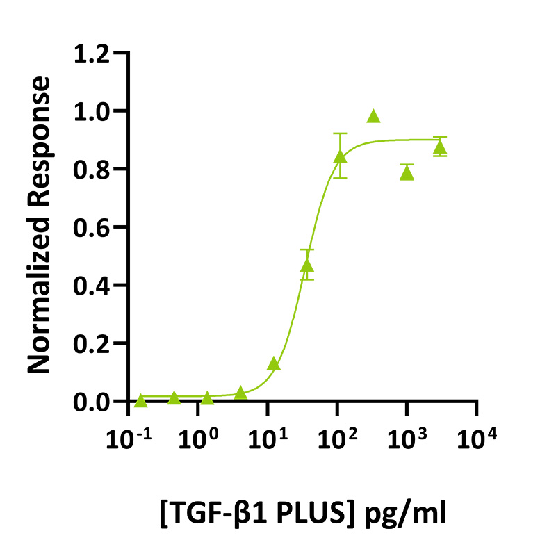
TGF-β1 PLUS™ activity was determined using the firefly luciferase reporter assay in transiently transfected HEK293T cells. Cells were treated in triplicate with a serial dilution of TGF-β1 PLUS™. Firefly luciferase activity was measured and normalized. EC50 = 34.8 pg/ml. Data from Qk010 lot #011.
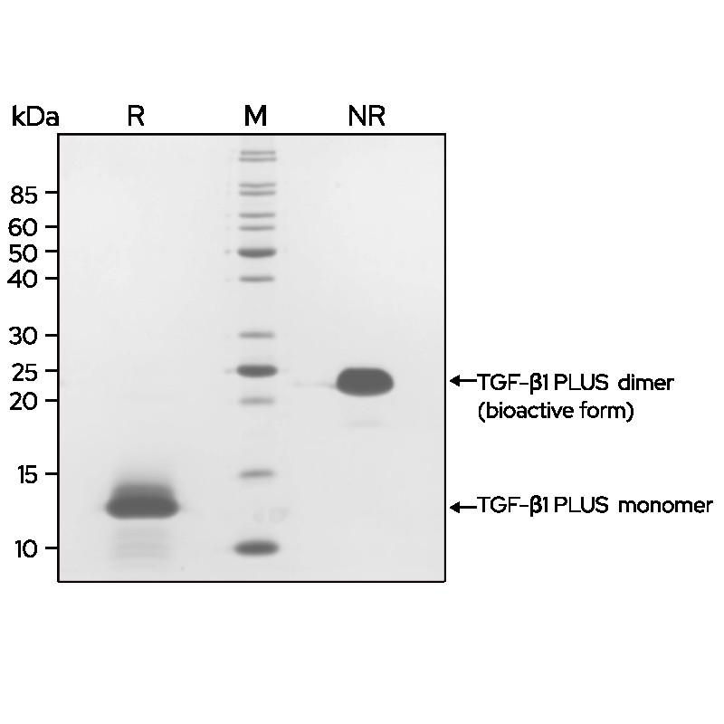
TGF-β1 PLUS™ (Qk010) dimer migrates as a single band at 24 kDa in non-reducing (NR) and 13 kDa as a single monomeric species upon reduction (R). High purity yield of dimeric protein (bioactive form). Purified recombinant protein (7 μg) was resolved using 15% w/v SDS-PAGE in reduced (+β-mercaptoethanol, R) and non-reduced conditions (NR) and stained with Coomassie Brilliant Blue R250. Data from Qk010 lot #012.
Mass spectrometry: single species with expected mass
Recovery from stock vial: >95%
Endotoxin: <0.05 EU/μg protein
We are a company founded and run by scientists to provide a service and support innovation in stem cell biology and regenerative medicine. All our products are exceptionally high purity, with complete characterisation and bioactivity analysis on every lot.

Qkine TGF-β1 PLUS™ was found to be more bioactive than mammalian-expressed TGF- β1 from alternative suppliers. Bioactivity was determined using a TGF-β1-responsive (CAGA) firefly luciferase reporter in transiently transfected HEK293T cells. Cells were treated with a serial dilution of TGF-β1 PLUS™ (Qk010, green) or TGF-β1 from two alternative suppliers (black) for 6 hours in triplicate. Firefly luciferase activity was measured and normalized to the control, Renilla luciferase.
Transforming growth factor-beta 1 (TGF-β1) is a pleiotropic cytokine part of the TGF-β superfamily. TGF-β1 regulates various cellular processes, including cell proliferation, growth, differentiation, motility, and apoptosis [1]. It plays a crucial role in the immune response, tissue repair, and the epithelial-mesenchymal transition. Transforming growth factor-beta 1 is produced by various cell types, including immune cells, fibroblasts, and epithelial cells. It is synthesized and secreted as an inactive or latent complex, associated with latency-associated proteins (LAP), and targeted to the extracellular matrix [2]. It is released from latency by TGF-β activators including plasmin, matrix metalloproteases, integrins. Once the LAP cleaved, the mature transforming growth factor-beta 1 is a homodimeric protein composed of two identical subunits linked by a disulfide bond. Its amino acid sequence is composed of 390 amino acids. TGF-β1 signals through complexes of cell surface receptors including TGF-βRII/TGF-βRI and ALK-5/ALK-1. This triggers downstream signaling cascades, such as the Smad-dependent and Smad-independent pathways.
In cell culture, recombinant TGF-β1 protein is an essential growth factor in many embryonic and induced pluripotent stem cell maintenance media, including the commonly used chemically-defined E8, StemPro, and mTeSR medias [3–5]. Transforming growth factor-beta 1 supports the survival and maintenance of pluripotency of stem cells [1]. TGF-β1 is used to promote the differentiation of various cell types such as fibroblasts, epithelial cells, and immune cells. It is used in combination with other growth factors such as BMP-2 to regulate bone marrow stromal cell differentiation or with IL-2 and IL-6 to regulate T reg and Th17 cells differentiation [6–8].
To date, recombinant human TGF-β1 has only been produced from mammalian cell protein expression systems (HEK or CHO), where endogenous protein contaminants, cost and animal-free status is a challenge. As part of the ongoing mission to redefine industry standards for growth factor and cytokine biochemical quality, Qkine introduced the first optimized, animal-free, and highly bioactive recombinant human TGF-β1.
Benefits:
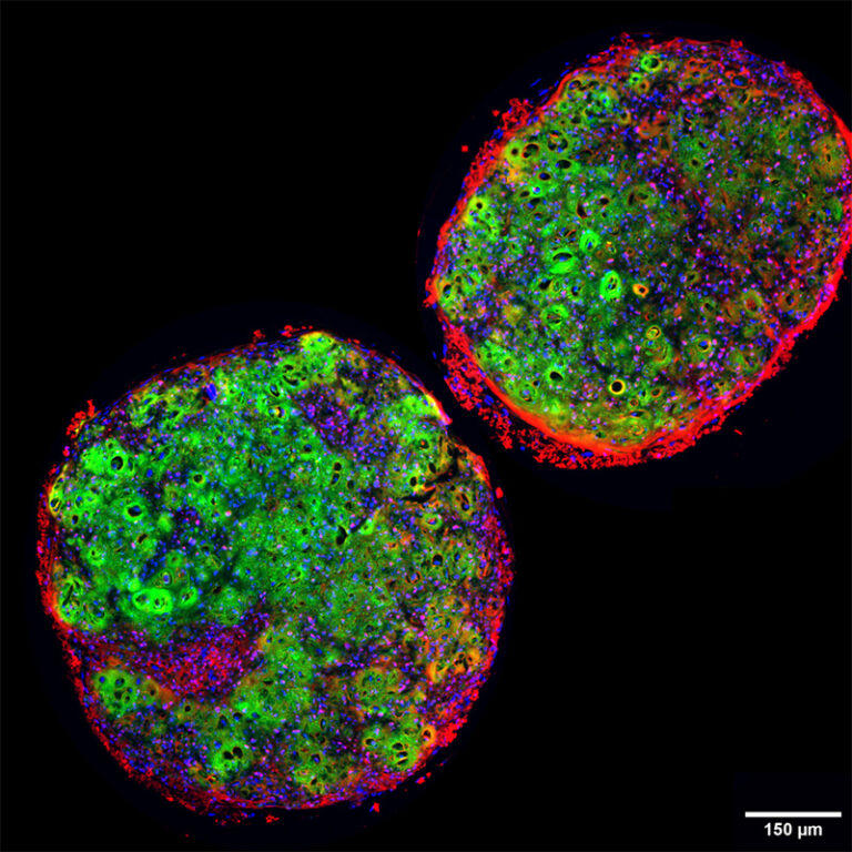
Cartilage spheroids made by using Qkine TGF-β1 PLUS™ (Qk010) to induce chondrogenic differentiation in iPSC derived mesodermal progenitor cells.
Data by Oliver Gardner/Valeriia Davydenko at Stem Cells and Regenerative Medicine, UCL.
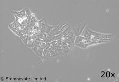
TGF-β1 PLUS™ (Qk010) maintains pluripotency and good colony morphology at 1 ng/ml in a chemically-defined serum and feeder-free iPSC culture. TGF-β1 PLUS™ used at 1 ng/ml; FGF-2 (Qk025) used at 100 ng/ml. Data from Stemnovate.
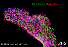
TGF-β1 PLUS™ is highly effective at maintaining iPSC pluripotency in chemically-defined, serum and feeder-free culture. Immuno-staining for pluripotency markers Tra 1-60 and Nanog show high levels of expression in iPSC lines maintained in TGF-β1 PLUS™ (Qk010)-containing defined media. In this study, Qkine TGF-β1 PLUS™ performed better than the TGF-β1 used routinely by Stemnovate.
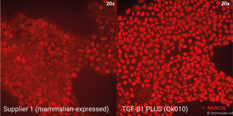
TGF-β1 PLUS™ in combination with Qkine FGF-2 shows enhanced bioactivity when benchmarked against other suppliers. Stemnovate IPS media (chemically-defined, serum and feeder-free culture). Higher nanog pluripotency marker immuno-staining in human iPSC cultured in Qkine FGF-2 (Qk025) and TGF-β1 PLUS™ (Qk010). TGF-β1 PLUS™ used at 1 ng/ml.
All experiments have been conducted by the specialist stem cell biotechnology company, Stemnovate Limited, in Cambridge, UK.
Beltran-Rendon C, Price CJ, Glen K et al.
DOI: doi: 10.1016/j.jcyt.2024.01.010Truszkowski L, Bottini S, Bianchi S et al.
DOI: doi.org/10.12688/openreseurope.18245.2Venkatesan M, Semper C, Skrivergaard S et al.
DOI: doi: 10.1016/j.isci.2022.105054.Rastovic U, Campinoti S, Wei L et al.
DOI: doi.org/10.1111/bph.70139Minerath RA, Kasam RK, Swoboda CO et al.
DOI: doi.org/10.1101/2025.09.22.677845Skowronska-Krawczyk et al.
DOI: doi.org/10.21203/rs.3.rs-8607320/v1Bottini S (Thesis)
DOI: ThesisHernanz M, Matas D, Mirasierra M et al.
DOI: DOI: 10.3791/69562Ng W, Aghakasiri K, Pierce R et al.
DOI: doi.org/10.1016/j.dib.2026.113042TGF-β1 is a multifunctional cytokine that plays a crucial role in various cellular processes. It regulates cell growth, differentiation, proliferation, migration, and apoptosis. It is also involved in the immune response, epithelial-mesenchymal transition, and the extracellular matric synthesis and modelling.
It is a member of the transforming growth factor-beta superfamily of cytokines that are synthesized as latent complexes. TGF-β1 requires further processing to become the biologically active, mature TGF-β1.
Dysregulation of TGF-β1 signaling is associated with various diseases such as cancer, fibrosis, autoimmune, cardiovascular, neurological, and chronic kidney diseases.
Elevated levels of TGF-β1 in the human body can have different implications such as higher risks of fibrosis, cancer progression, chronic inflammation, and cardiovascular diseases. It is important to consider the specific clinical context, the presence of other biomarkers, and the patient’s prognosis.
TGF-β1 is synthesized and secreted in an inactive or latent form. It is activated through cleavage and binding to matrix metalloproteinases, integrins, extracellular matrix such as thrombospondin-1, but also pH changes and reactive oxygen species.
It is an essential growth factor in many embryonic and induced pluripotent stem cell maintenance media, including the commonly used E8, StemPro, and mTeSR media. TGF-β1 also promotes the differentiation of various cell types such as fibroblasts, epithelial cells, and immune cells.
Qkine TGF-β1 PLUS™ protein is the first entirely animal origin-free recombinant TGF-β1 protein, TGF-β family members are difficult to express in E. coli due to their complex folding so are only commercially available expressed in animal cells. Qkine have optimized the production of TGF-β1 to produce a pure animal origin-free protein or highly reproducible results and compatible with chemically-defined stem cell media.
TGF beta family proteins and other growth factors can be very poorly soluble in physiological solutions. Please follow the handling guidance for lyophilized cytokines below to minimize loss of protein due to precipitation or adsorption to plastic. We advise storing the recombinant protein at very low pH before dilution in cell culture media or final working solutions. Low pH will also assist in maintaining the correct disulfide structure of the protein by minimizing disulfide bond exchange reactions.
Every effort is made to ensure samples are sterile; however, we recommend sterile filtering after dilution in media or the final working solution.
Our products are for research use only and not for diagnostic or therapeutic use. Products are not for resale.
For use in manufacturing of cellular or gene therapy products. Not intended for in vivo applications.

£100, $140 or €120 Qkine gift card for product reviews with an image and £50, $70 or €60 for reviews without an image.
Recombinant human TGF-β1 PLUS™ protein (Qk010)