Quality commitment 4
Subject to quantitative bioactivity analysis with integrated lot-to-lot reproducibility criteria
At Qkine we test all our growth factors and cytokines for bioactivity, each new lot is validated for bioactivity comparable to previous lots before they are released into the supply chain.
Validated assays for reliable bioactivity testing
At Qkine our in-house R&D team test all our cytokines and growth factors to ensure their bioactivity.
Protein-specific quantitative luciferase reporter assays to determine signaling pathway activation.
Proliferation assays to accurately determine stimulation of cell proliferation.

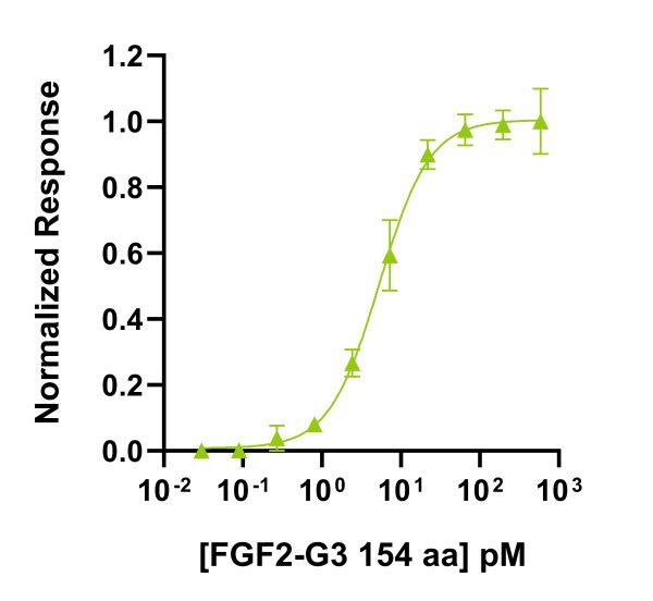
Recombinant FGF2-G3 (Qk053) activity was determined using the Promega serum response element luciferase reporter assay (*) in transfected HEK293T cells. Cells were treated in triplicate with a serial dilution of FGF2-G3 for 3 hours (EC50 = 5.2 pM). Data from Qk053 lot #104340. *Promega pGL4.33[luc2P/SRE/Hygro] #E1340
Lot-to-lot validation of bioactivity
Qkine test the bioactivity of each new lot produced vs previous lots for consistency in bioactivity in supplied proteins. Lots which are significantly different to previous lots are disposed of and not moved into the supply chain.

Bioactivity of Qkine BMP-4 (Qk038) was determined using a BMP-4-responsive firefly luciferase reporter assay in stably transfected HEK293T cells. Cells were treated with a serial dilution of four independent lots of BMP-4 for 6 hours in triplicate. See BMP-4 technote.
Bioactivity compared with alternative supplier proteins
As part of our Bioactivity Guarantee, we conduct comparative quantitative bioactivity studies with current dominant suppliers to ensure the bioactivity of our proteins is equivalent or better. During our R&D process we always correct for protein concentration after reconstitution, to ensure we are comparing like-for-like and correct for any discrepancies in vial recovery.

Quantitative luciferase reporter assay shows equivalent bioactivity of IL-6 (Qk093, green) and alternative supplier IL-6 (Supplier B, black). HEK293T luciferase reporter cells were treated in triplicate with a serial dilution of IL-6 for 24 hours. Firefly luciferase activity is measured and normalized to control Renilla luciferase activity. See IL-6 technote.
Getting the simple things right is important.
At Qkine, we are committed to raising the standard in bioactive protein manufacturing.
Our science team is here to help, please contact us if you have any questions at customerservice@qkine.com.
Quantitative Luciferase Assay
It can be hard to determine a single value for the activity of a growth factor as growth factors generally have a large range of potential effects. A luciferase reporter assay is used to quantify the response of transfected cells to a growth factor by assaying the expression of luciferase (the reporter gene). As the biological activity of a growth factor is measured by its effect on a defined cell type in standardized conditions, it may not accurately represent your own culture conditions, however, it will provide confirmation that the growth factor is able to elicit a biological response.
To determine this bioactivity, a vector containing a firefly luciferase gene under the control of a growth factor-appropriate regulatory region is introduced into cells, such as HEK293Ts. These are then incubated with a dilution series of the growth factor. After a defined incubation period, the bioactivity is measured by quantifying the enzymatic activity of luciferase. This involves lysing the cells and adding the luciferase substrate, luciferin. The amount of light emitted from the reaction is directly proportional to the amount of luciferase in the sample and consequently can be used as a measure of the level of response the growth factor imparts on the reporter gene. In our QC experiments, we also quantify Renilla luciferase which is expressed constitutively by co-transfecting the same cells with a vector containing a Renilla luciferase gene. This acts as a normalization control for transfection efficiency and the cell number in each well. The growth factor response can then be expressed as a firefly/Renilla ratio. Plotting concentration vs. F/R ratio allows an EC50 (half-maximal effective concentration) to be calculated.
Proliferation Assays
The bioactivity of some of our growth factors is measured using a proliferation assay. The ability of the growth factor to stimulate the proliferation of a relevant cell type is measured through a luminescence method related to the number of live cells (amount of ATP) after a defined number of days in culture. Again, plotting growth factor concentration vs. luciferase units allows an EC50 to be calculated.
The EC50 is the concentration of the growth factor which gives 50% of the maximum effect on the given cell population.
Raising the standard in bioactive protein manufacturing and innovation
Our science team is here to help, please contact us if you have any questions.