 Recombinant human IL-1β protein (Qk101)
 Price range: £320.00 through £3,960.00View product
Price range: £320.00 through £3,960.00

Interleukin-1 beta (IL-1β) is a pro-inflammatory cytokine and a potent mediator of inflammation and immune responses. It promotes the activation and recruitment of immune cells to sites of infection or injury, such as antigen presenting cells, macrophages, and T lymphocytes. In cell culture, recombinant IL-1β is used to stimulate and activate immune cells (macrophages and T cells), maintain hematopoietic progenitor cells, and modulate the differentiation of mesenchymal stem cells.
Qkine human IL-1β is a monomer of a molecular weight of 17.3 kDa. This protein is animal origin-free, carrier protein-free, and tag-free to ensure a homogenous population with exceptional lot-to-lot consistency. IL-1β is suitable for reproducible, high-quality hematopoietic stem cells and more specific lineages.
This protein is also available as GMP compliant Cell Therapy Grade, to enquire email [email protected].
In stock
Orders are typically shipped same or next day (except Friday).
Easy world-wide ordering, direct or through our distributors.
Price range: £320.00 through £3,960.00
Buy online with secure credit card or purchase order. For any questions, please email [email protected]
>98%, by SDS-PAGE quantitative densitometry
Expressed in E. coli
Animal origin-free (AOF) and carrier protein-free
Manufactured in our Cambridge, UK laboratories
Lyophilized from HEPES, NaCl, Cys
Maintenance and differentiation of hematopoietic stem cells
Directed osteogenic differentiation of mesenchymal stem cells
Neural stem cell proliferation and neuronal differentiation
Activation of M2 macrophages
Activation of Th17 cells
IL-1β activity was determined using the IL-1β-responsive firefly luciferase reporter assay. Transfected HEK293T cells were treated in triplicate with a serial dilution of IL-1β for 3 hours. Firefly luciferase activity was measured and normalised to the control Renilla luciferase activity. Data from Qk101 lot #204587. EC50 = 71 pg/ml (4 pM).
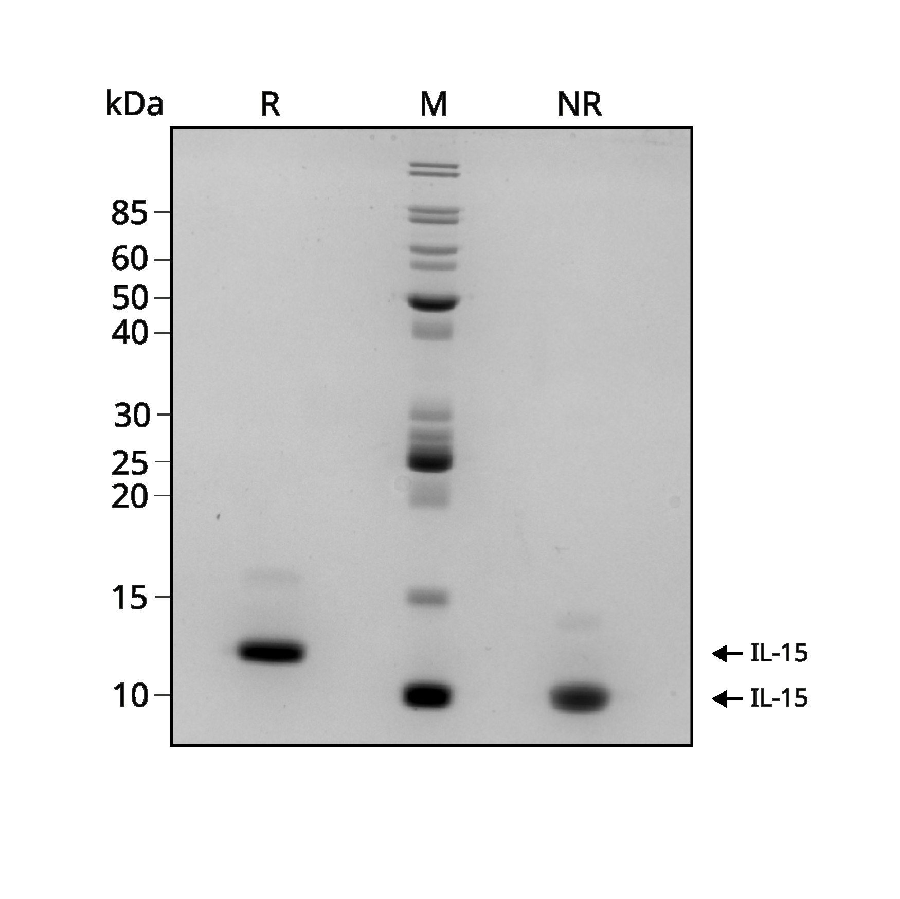
Recombinant IL-1β migrates as a major band at 17.4 kDa in non-reducing (NR) conditions. The higher molecular weight minor band is the dimeric form. Upon reduction (R), only the 17.4 kDa band is visible. No contaminating protein bands are present. Purified recombinant protein (3 µg) was resolved using 15% w/v SDS-PAGE in reduced (+β-mercaptothanol, R) and non-reduced (NR) conditions and stained with Coomassie Brilliant Blue R250. Data from Qk101 batch #204587.

Mass spectrometry: single species with expected mass
Recovery from stock vial: >95%
Endotoxin: <0.05 EU/μg protein
We are a company founded and run by scientists to provide a service and support innovation in stem cell biology and regenerative medicine. All our products are exceptionally high purity, with complete characterisation and bioactivity analysis on every lot.

Qkine IL-1 beta is slightly more biologically active than a comparable alternative supplier protein. Bioactivity was determined using a NFkB quantitative luciferase assay. HEK293T luciferase reporter cells were treated in triplicate with a serial dilution of Qkine IL-1 beta (Qk101, green) or an alternative supplier (supplier B, black) for 3 hours. Firefly luciferase activity is measured and normalized to control Renilla luciferase activity. Data from Qk101 lot #204581.
Interleukin-1 beta (IL-1β) is a pro-inflammatory cytokine and a member of the interleukin-1 family, including IL-1α [1]–[4]. IL-1β is a potent mediator of inflammation and immune responses. It plays a key role in initiating and amplifying the inflammatory cascade in response to infection, injury, or other pathological conditions [2], [5], [6]. It promotes the activation and recruitment of immune cells, such as antigen-presenting cells, macrophages, and T lymphocytes, to sites of infection or injury and induces fever [3], [5]. IL-1β also stimulates the production of acute-phase proteins in the liver, contributing to systemic responses during inflammation, such as blood clotting and tissue repair [1], [7].
In cell culture, IL-1 beta is used to stimulate various immune cells (macrophages, monocytes, and T cells) as well as epithelial cells, endothelial cells and fibroblasts [1], [8]. IL-1β inhibits the B cell differentiation and promotes the proliferation, survival, and polarization of macrophages towards the M2 phenotype and CD4+ T cells towards T helper type 1 and 17 (Th1 and Th17) cells [5], [9], [10]. IL-1β mimics the effects of inflammation along with tumor necrosis factor-alpha (TNF-α) and promotes the release of pro-inflammatory cytokines and other inflammatory mediators [2], [11]. IL-1 beta is also used to maintain mesenchymal stromal cells and hematopoietic progenitor cells and to modulate the differentiation of mesenchymal stem cells into osteoblasts, neural stem cells into neurons and astrocytes, and endothelial cells [12], [13].
IL-1β is produced primarily by activated macrophages and monocytes, as well as epithelial cells, endothelial cells, and specific fibroblasts, in response to various stimuli such as infection, tissue damage, or stress. IL-1β is a protein composed of 153 amino acids. It is synthesized as an inactive precursor, pro-IL-1β, which requires cleavage by the enzyme caspase-1 to become active [1]. The activity of IL-1β is tightly regulated to prevent excessive inflammation. In addition to the cleavage of pro-IL-1β by caspase-1, other regulatory mechanisms, such as IL-1 receptor antagonist (IL-1Ra) and soluble receptors, modulate IL-1β signaling [6]. IL-1β is characterized by a beta-trefoil fold, six antiparallel beta-strands arranged in a threefold symmetric pattern. IL-1 beta binds to specific cell surface receptors, primarily the IL-1 receptor type 1 (IL-1R1) and IL-1 receptor accessory protein (IL-1RAcP) [5], [14]. This binding triggers a signaling cascade, leading to the activation of transcription factors and the expression of pro-inflammatory genes.
Dysregulation of IL-1β is associated with various chronic inflammatory diseases, including autoimmune diseases, rheumatoid arthritis, type 2 diabetes, and inflammatory bowel disease [5], [15]. IL-1β may also support the tumor development by increasing tumor-associated macrophages and sustaining chronic inflammation and metastasis [5]. Therapeutic interventions targeting IL-1β, such as IL-1β inhibitors, have been developed for the treatment of inflammatory disorders.
Our products are for research use only and not for diagnostic or therapeutic use. Products are not for resale.
For use in manufacturing of cellular or gene therapy products. Not intended for in vivo applications.

£100, $140 or €120 Qkine gift card for product reviews with an image and £50, $70 or €60 for reviews without an image.
Recombinant human IL-1β protein (Qk101)