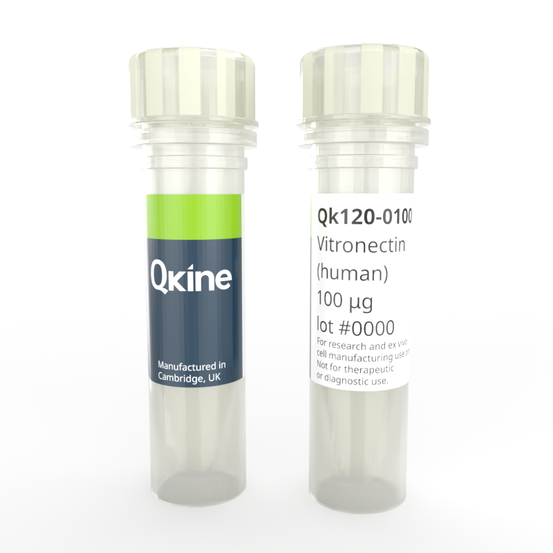
 Recombinant human vitronectin protein (Qk120)
Recombinant human vitronectin protein (Qk120)
Price range: £110.00 through £460.00
Vitronectin protein is widely used in stem cell culture. It provides a defined environment that supports the maintenance of pluripotency and is suitable for feeder-free culture, expansion, differentiation, and reprogramming of stem cells.
Qkine vitronectin is a high-purity animal origin-free recombinant protein with a molecular weight of 47.8 kDa. It is carrier-free and protein tag-free, ensuring exceptional lot-to-lot consistency and compatibility with translational studies and iPSC-based model production for screening. Qkine recombinant vitronectin protein is ultra high-purity with exceptionally low residual endotoxins making it suitable for the reproducible culture of stem cells, primary cells, organoids, and use in sensitive neural differentiations.
In stock
Orders are typically shipped same or next day (except Friday).
Easy world-wide ordering, direct or through our distributors.
Price range: £110.00 through £460.00
Buy online with secure credit card or purchase order. For any questions, please email orders@qkine.com
Summary:
- Highly pure recombinant human vitronectin protein (truncated) (UniProt: P04004)
- 47.8 kDa (monomer)
>98%, by SDS-PAGE quantitative densitometry
Expressed in E. coli
Animal origin-free (AOF) and carrier protein-free
Manufactured in our Cambridge, UK laboratories
Lyophilized from PBS, mannitol and TCEP
- Resuspend in sterile-filtered water at 1 mg/ml, add carrier protein if desired, prepare single use aliquots and store frozen at -20 °C (short-term) or -80 °C (long-term).
Featured applications:
Induced pluripotent and embryonic stem cell differentiation and maintenance
Differentiation of human pluripotent stem cells towards extra-embryonic endoderm, mesenchymal, neural lineages, and chondrocytes
Promotion of cell adhesion and migration
Organoid growth and proliferation
Stimulation of angiogenesis and vascular network development
Fully defined, animal-free protocols for translational studies and preclinical stem cell production

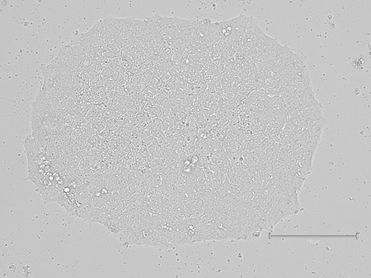
Imaging of iPSC colonies grown on Qkine vitronectin coated plates in E8-like media retained their highly preserved morphological appearance. (A) after initial seeding and 3 days in culture; (B) After 1 passage and 7 days in culture; (C) After 3 passages and 14 days in culture (scale bar = 150 µm). 6-well plates were coated with 5 µg/ml Qk120 vitronectin protein lot #204660.
Preparation of cell culture plates with Qkine ultra-high quality vitronectin (Qk120)
- Briefly centrifuge vial containing vitronectin Qk120 to ensure all lyophilized protein is collected at the bottom of the vial.
- Resuspend in 500 ml of MilliQ water to make a 1 mg/ml stock and lightly agitate the tube to ensure everything has fully reconstituted.
- Per 6-well plate required, dilute 30 µl of the 1 mg/ml in 6 ml of phosphate buffered saline (PBS) without MgCl and CaCl to make 5 µg/ml solution.
- Coat each well of 6-well plate with 1 ml per well of 5 µg/ml vitronectin for at least two hours at 37°C.
- Adjust volume of 5 µg/ml vitronectin per well for different area plate clusters.
- 12-well plate add 500 µl
- 24-well plate add 250 µl
- 96-well plate add 100 µl
- Aliquot the remaining resuspended 1 mg/ml vitronectin into appropriately sized single use aliquots and store at -80°C.
Further quality assays
Mass spectrometry: single species with expected mass
Recovery from stock vial: >95%
Endotoxin: <0.005 EU/μg protein (below level of detection)
We are a company founded and run by scientists to provide a service and support innovation in stem cell biology and regenerative medicine. All our products are exceptionally high purity, with complete characterisation and bioactivity analysis on every lot.
Protein background
Vitronectin is a glycoprotein found in the extracellular matrix (ECM) and blood plasma. It plays a crucial role in various physiological processes, including cell adhesion, migration, wound healing, and immune response regulation [1]. Vitronectin interacts with various cell surface receptors and other ECM proteins, making it a key player in tissue remodeling and repair [2].
Vitronectin is involved in numerous biological processes, including cell adhesion and migration by binding to integrins, wound healing and tissue repair, regulation of proteolysis and fibrinolysis, immune response regulation, and angiogenesis by promoting the formation of new blood vessels and supporting tumorigenesis and metastasis [3-5].
In cell culture, vitronectin has been widely used due to its key roles in cell adhesion, proliferation, differentiation, and survival. It interacts with integrins and other cell surface receptors, creating a physiologically relevant microenvironment for various cell types, particularly in stem cell research and regenerative medicine [6-7]. As a coating substrate, vitronectin promotes cell attachment, especially for epithelial cells, endothelial cells, and certain cancer cells. By binding to integrins, vitronectin enhances cell proliferation and survival through the activation of intracellular signaling pathways such as PI3K/AKT and MAPK. This attachment prevents anoikis, a form of cell death that occurs when cells detach from the ECM, ensuring the maintenance and expansion of cultured cells [8].
In stem cell applications, vitronectin is used for the culture and maintenance of iPSCs and ESCs, promoting their efficient differentiation into various lineages. It is also a key component of defined, xeno-free culture systems, eliminating the need for animal-derived products, reducing variability, and improving reproducibility in stem cell research [7,9-11].
In organoids technology, vitronectin plays a critical role in the development and maintenance of organoids due to its properties that enhance cell adhesion, proliferation, and differentiation. As a matrix scaffold, vitronectin supports the initial adhesion and aggregation of cells, essential for stable organoid structure formation [12] By binding to integrins such as αvβ3 and αvβ5 on cell surfaces, vitronectin facilitates cell clustering, a prerequisite for organoid growth [13] It activates integrin-mediated signaling pathways like PI3K/AKT and MAPK/ERK, promoting cell survival, proliferation, and differentiation, which are crucial for expanding progenitor cells and their differentiation within the organoid. Vitronectin has been implicated in the development of liver organoid [14].
In tissue engineering and regenerative medicine, vitronectin-coated scaffolds and biomaterials enhance cell attachment, proliferation, and differentiation, contributing to the development of functional tissue constructs [15-16]. Additionally, vitronectin is used to prepare cells for cell therapy, ensuring their viability and functionality upon transplantation.
Vitronectin is particularly important for optimizing culture conditions in the manufacturing of serum-free media, compensating for the lack of adhesion-promoting factors typically provided by serum and supporting cell growth under defined conditions [7, 17]. The use of recombinant vitronectin ensures batch-to-batch consistency, which is critical for reproducibility in experimental and clinical applications.
Structurally, vitronectin is a multifunctional glycoprotein with a molecular weight of approximately 75 kDa. It comprises several domains, including the somatomedin B domain at the N-terminal, which is important for binding to the urokinase receptor and stabilizing the inactive form of plasminogen activator inhibitor-1 (PAI-1). The RCL (Ricinus communis agglutinin-like) domain is crucial for heparin binding, while the hemopexin-like repeats facilitate interactions with integrins and other ECM components [2,6,18]. Vitronectin can undergo conformational changes that expose or hide specific binding sites, enabling it to interact with a wide range of ligands and receptors.
In summary, vitronectin plays a crucial role in various applications, particularly in stem cell and organoid applications, by providing a defined and supportive environment for stem cell maintenance, differentiation, and reprogramming. Its use enhances the reproducibility and efficiency of several cell cultures, making it a valuable tool in both research and therapeutic contexts.
Additional resources
- Technote | Human vitronectin (Qk120) coating
- Differentiation of induced pluripotent stem cells (iPSCs) into neuroectoderm (PDF)
- Differentiation of induced pluripotent stem cells (iPSCs) into mesoderm (PDF)
- Weekend-free human induced pluripotent stem cell culture using thermostable FGF-2 (bFGF) and animal origin-free TGF-β1 and vitronectin for improved colony homogeneity (PDF)
- Differentiation of induced pluripotent stem cells (iPSCs) into microglia
FAQ
A glycoprotein found in the extracellular matrix (ECM) that plays critical roles in cell adhesion, migration, wound healing and regulation of immune response.
Vitronectin is found in the extracellular matrix, blood plasma and various tissues.
Vitronectin is not a cytokine but it signals through integrin receptors to support maintenance of pluripotency during iPSC culture.
Vitronectin gene encodes vitronectin protein, which is a component of the extracellular matrix.
Vitronectin binds to integrins, heparin and heparan sulfate proteoglycans, plasminogen activator inhibitor – 1, complement system.
The vitronectin receptor facilitates cell adhesion, cell migration and initiates signaling processes that promotes cell survival, proliferation and differentiation.
Vitronectin is widely used in stem cell culture. It provides a defined environment than supports maintenance of pluripotency and is suitable for feeder-free culture, maintenance, differentiation, and reprogramming of stem cells.
Our products are for research use only and not for diagnostic or therapeutic use. Products are not for resale.
For use in manufacturing of cellular or gene therapy products. Not intended for in vivo applications.

Receive an Amazon gift voucher when you leave us a review.
£25, $30 or €30 for reviews with an image and £10, $15 or €15 for reviews without an image

